Thyroidectomy can range from a straightforward procedure to one that is fairly complex. Benign, well-encapsulated tumors, such as those found in most cats, are easily resectable with minimal complications. Malignant, invasive tumors require extensive, careful dissection around many important and vital structures such as the trachea, esophagus, carotid arteries, jugular veins, and recurrent laryngeal nerves (3-6).
A good working knowledge of the regional anatomy of the thyroid and parathyroid glands, as well as proper pre- and postoperative care is necessary for successful patient management with thyroidectomy (3-7).
The purpose of this post is to provide an overview of the different surgical techniques for thyroidectomy in the cat.
Positioning of the Hyperthyroid Cat for Thyroidectomy
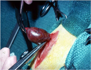
Thyroidectomy in the cat is performed by a ventral midline (neck) cervical approach (Figure 1).
 |
| Figure 1: Preparation and positioning of a cat for thyroidectomy |
Towel clamps should penetrate only through the skin to avoid trauma to the jugular veins. Bipolar cautery, fine scissors and thumb forceps, sterile cotton-tipped applicator swabs, and magnification are useful during dissection of the parathyroid glands and removal of a cat’s thyroid tumors.
Initial Surgical Exploration
After proper positioning of the cat (see Figure 1), the 2 thyroid lobes are exposed through a ventral midline cervical approach. To accomplish this, a skin incision is first made from the larynx to the upper part of the sternum (manubrium); the incision is then continued through the subcutaneous tissues and superficial oblique muscle fibers (3-6). The paired sternohyoideus muscles are then exposed and separated in the midline to expose the trachea. Care is taken to avoid cutting blood vessels that lie on the ventral surface of the trachea.
Identify both thyroid lobes
The two thyroid lobes are next located (Figure 2). Normally, both lobes are located just caudal (posterior) to the larynx (voice box) on the medial (inside) aspect of the sternothyroideus muscles. However, an adenomatous thyroid lobe can be located anywhere between the larynx and thoracic inlet, as gravity pulls the thyroid tumor down ventrally in the neck.
 |
| Figure 2: Locating both thyroid lobes in a hyperthyroid cat with bilateral thyroid disease |
Identify external parathyroid glands
 |
| Figure 3: Parathyroid gland anatomy |
The parathyroid glands can sometimes be difficult to differentiate from fat deposits on the surface of the thyroid capsule. Under magnification, each parathyroid gland will have a small vessel that splits and surrounds the gland.
Surgical Techniques for Thyroidectomy
Four different techniques have been described for performing a thyroidectomy in cats with hyperthyroidism (3-6). These include the following:
- Extracapsular thyroidectomy technique
- Modified extracapsular technique
- Intracapsular thyroidectomy technique
- Modified intracapsular technique
The aim of all of these thyroidectomy techniques is to remove all abnormal thyroid tissue and preserve at least 1 parathyroid gland. In addition, care are should be taken to avoid trauma to the adjacent vessels and nerves, as well as to the parathyroid glands.
 |
| Figure 4: Performing a unilateral thyroidectomy in a cat with the extracapsular technique |
References:
- Mooney CT, Peterson ME: Feline hyperthyroidism, In: Mooney C.T., Peterson M.E. (eds), Manual of Canine and Feline Endocrinology (Fourth Ed), Quedgeley, Gloucester, British Small Animal Veterinary Association, 2012; 199-203.
- Baral R, Peterson ME: Thyroid gland disorders, In: Little, S.E. (ed), The Cat: Clinical Medicine and Management. Philadelphia, Elsevier Saunders. 2012; 571-592.
- Birchard, SJ. Thyroidectomy in the cat. Clinical Techniques in Small Animal Practice 2006;21:29-33.
- Flanders JA. Surgical therapy of the thyroid. Veterinary Clinics of North America. Small Animal Practice 1994;24:607–621.
- Padgett S. Feline thyroid surgery. Veterinary Clinics of North America. Small Animal Practice 2002;32:851–859.
- Panciera DL, Peterson ME, Birchard, SJ: Diseases of the thyroid gland. In: Birchard SJ, Sherding RG (eds): Manual of Small Animal Practice (Third Edition), Philadelphia, Saunders Elsevier, pp 327-342, 2006.
- Waters DJ. Endocrine system. In: Hudson LC, Hamilton WP (eds): Atlas of Feline Anatomy for Veterinarians. Philadelphia: WB Saunders, 1993;127–134.


No comments:
Post a Comment