As discussed in my last post we have several different techniques that can be used when performing a thyroidectomy in cats with hyperthyroidism (1-8). These include the following:
- Extracapsular thyroidectomy technique
- Modified extracapsular technique
- Intracapsular thyroidectomy technique
- Modified intracapsular technique
The aim of all of these thyroidectomy techniques is to remove all abnormal thyroid tissue and preserve at least 1 parathyroid gland. In addition, care are should be taken to avoid trauma to the adjacent vessels and nerves, as well as to the parathyroid glands.
The 4 Surgical Techniques for Thyroidectomy in Cats
Extracapsular technique
The “original” extracapsular technique (1) is most useful for cats with unilateral thyroid disease, in which only one thyroid lobe needs to be removed. With this technique, the affected thyroid tumor together with the associated external and internal parathyroid glands are removed.
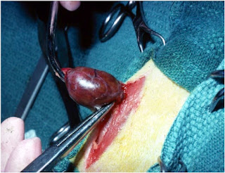
This surgical procedure here is simple: once the thyroid tumor is identified, the cranial and caudal blood supply to the affected thyroid tumor is ligated, and the entire thyroid lobe is excised along with its capsule (Figure 1). Again, no attempt is made to preserve the external parathyroid gland with this method (1,2).
 |
| Figure 1: Performing a unilateral thyroidectomy in a cat with the extracapsular technique |
Because the entire thyroid tumor and its capsule are removed with this technique, the cure rate is high, with little chance of local recurrence. However, because the external parathyroid gland is also removed, this technique is not recommended for cats in which bilateral thyroidectomy is needed because of the very high incidence of hypoparathyroidism (4,5,9).
Modified extracapsular technique
The “modified” extracapsular technique was developed to decrease the risk of postoperative hypoparathyroidism and hypocalcemia that can develop when both thyroid lobes are removed in cats with bilateral thyroid adenomas (3,5,7,8).
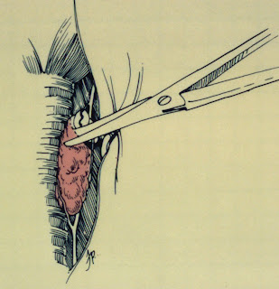
Compared to the original extracapsular technique, this surgical procedure is more difficult. Once the affected thyroid tumors and external parathyroid glands are identified, the thyroid gland capsule is incised approximately 300 degrees around the external parathyroid gland (Figure 2), being careful to preserve the blood supply to the parathyroid gland.
 |
| Figure 2: Extracapsular dissection for removal of a thyroid tumor in a cat. Figure from reference 8 (with permission) |
With the modified extracapsular technique, the thyroid tumor and approximately 90% of its thyroid capsule are removed, leaving a small rim of thyroid capsule around the external parathyroid gland (3,5,7,8). Use of this modified extracapsular technique helps ensure that the external parathyroid gland with its blood supply remain intact, greatly lessening the incidence of hypoparathyroidism. However, because a small amount of thyroid capsule is not removed, small remnants of adenomatous tissue that are left behind may regrow with time, leading to recurrence.
Intracapsular technique
With the intracapsular technique for thyroidectomy, a small nick incision is made in an avascular area of the thyroid capsule on the middle to caudal aspect of the gland (Figure 3A). This longitudinal incision is extended with a scalpel blade or fine iris scissors until the entire thyroid capsule is opened (Figure 3B). The incised thyroid capsule is reflected off the gland with tissue forceps (Figure 3C). The thyroid tumor tissue is then gently teased away from the inside aspect of the thyroid capsule with a sterile cotton-tipped applicator, leaving the thyroid capsule and external parathyroid gland intact.
 |
| Figure 3: Intracapsular dissection for removal of a thyroid tumor in a cat. Figure from reference 8 (with permission) |
The advantage of the intracapsular technique over the extracapsular techniques, described above, are that this is a technically simple method to help ensure that the external parathyroid gland and delicate blood supply are preserved. Because of this, the risk of hypoparathyroidism is greatly reduced. However, because of retained remnants of abnormal thyroid tissue that remain attached to the thyroid capsule, the rate of recurrent hyperthyroidism is highest with this method (4,10,11).
Modified intracapsular technique
Because the intracapsular technique has the potential to leave a significant amount of thyroid tumor tissue behind, a “modified” intracapsular technique was subsequently developed for use in hyperthyroid cats.
The procedure is as described above for the intracapsular technique, but as a final step, the thyroid capsule (caudal to the parathyroid gland) is resected following removal of the thyroid tumor. This leaves only a small rim of thyroid capsule around the external parathyroid gland, thereby greatly lessening the chance of recurrence (3,7).
Staged Bilateral Thyroidectomy
Some surgeons recommend that bilateral thyroidectomy be performed in two stages or separate surgical procedures, in order to lessen the risk of hypoparathyroidism (4). A period of at least 3-4 weeks between procedures gives time for vascular or parathyroid damage to heal.
The necessity of two anesthetic episodes is the major drawback of the technique, considering the older age of the hyperthyroid cats often affected.
Parathyroid Gland Autotransplantation
Parathyroid autotransplantation has also been described as a treatment for accidental removal of the parathyroid or if complete devascularization occurs during thyroidectomy (7,9).
If the parathyroid glands are removed or damaged, the parathyroid gland can be minced into small 1-mm pieces and inserted into a small pocket made in the cervical musculature. With time, such transplanted parathyroid tissue can start to function again. This will decrease the severity and duration of postoperative hypocalcemia.
My Bottom Line
I greatly prefer the extracapsular technique for thyroidectomy in cats because it helps to ensure a permanent cure of the cat’s hyperthyroidism (i.e., the recurrence rate is extremely low). With the intracapsular techniques (especially the original technique), remnants of thyroid tissue are commonly left behind, which can regrow to cause recurrence of hyperthyroidism in some cats.
However, in cats in which the parathyroid glands can not be identified, we still rely on intracapsular dissection when an external parathyroid gland cannot be identified. With this technique, we open the thyroid capsule, which makes it easier to locate and preserve at least one of the parathyroid glands and prevent hypoparathyroidism.
Finally, in cats in which identifying or saving the parathyroid gland is difficult, use of the staged bilateral thyroidectomy approach, parathyroid gland transplantation, or both can help lessen the incidence of postoperative hypocalcemia. However, considering the increased cost and morbidity of a second operation, our preference is to perform bilateral thyroidectomy in one procedure if at all possible.
References:
- Black AP, Peterson ME: Thyroid biopsy and thyroidectomy, In: Bojrab MJ (ed): Current Techniques in Small Animal Surgery. Philadelphia, Lea and Febiger. 1983; 388-396.
- Birchard SJ, Peterson ME, Jacobson A. Surgical treatment of feline hyperthyroidism: Results of 85 cases. Journal of the American Animal Hospital Association 1984;20:705-709.
- Birchard, SJ. Thyroidectomy in the cat. Clinical Techniques in Small Animal Practice 2006;21:29-33.
- Flanders JA, Harvey HJ, Erb HN. Feline thyroidectomy. A comparison of postoperative hypocalcemia associated with three different surgical techniques. Veterinary Surgery 1987;16:362–366.
- Flanders JA. Surgical therapy of the thyroid. Veterinary Clinics of North America. Small Animal Practice 1994;24:607–621.
- Flanders JA. Surgical options for the treatment of hyperthyroidism in the cat. Journal of Feline Medicine and Surgery 1999;1:127–134.
- Padgett S. Feline thyroid surgery. Veterinary Clinics of North America. Small Animal Practice 2002;32:851–859.
- Panciera DL, Peterson ME, Birchard, SJ: Diseases of the thyroid gland. In: Birchard SJ, Sherding RG (eds): Manual of Small Animal Practice (Third Edition), Philadelphia, Saunders Elsevier, pp 327-342, 2006.
- Padgett SL, Tobias KM, Leathers CW, et al. Efficacy of parathyroid gland autotransplantation in maintaining serum calcium concentrations after bilateral thyroparathyroidectomy in cats. Journal of the American Animal Hospital Association 1998;34:219-224.
- Swalec KM , Birchard SJ. Recurrence of hyperthyroidism after thyroidectomy in cats. Journal of the American Animal Hospital Association. 1990;26:433–437.
- Welches CD, Scavelli TD, Matthiesen DT, Peterson ME. Occurrence of problems after three techniques of bilateral thyroidectomy in cats. Veterinary Surgery 1989;18:392-396.


4 comments:
Hi,
this is a question about thyroid levels in a cat.
We took our approx 9-10yr old cat in for bloodwork last week, both to re-check her liver values and for a comprehensive pre-dental work-up. She was found to be hyperthyroid; T4: 4.1, free T4: 74.
However, in August of this year her T4 was 0.6! (In November 2011 it was 2.2, in January 2011 it was 1.8.) What could cause her temporarily and dramatically low T4 level? (I do not have free T4 values for any of these dates.)
We don't know the cause of her liver value elevations, here is the history in case it is relevant:
8/30/12
ALT 124 [10-100] HIGH
AST 43 [10-100] normal
Alk Phosphatase 194 [6-102] HIGH
Triglycerides 174 [25-160] HIGH
10/9/2012
ALT 70 [10-100] normal
AST not tested
Alk Phosphatase 176 [6-102] HIGH
Triglycerides not tested
11/23/12
ALT 115 [10-100] HIGH
AST 45 [10-100] normal
Alk Phosphatase 173 [6-102] HIGH
Triglycerides 80 [25-160] normal
CPK 1916 [56-529] HIGH
She is on Denosyl and milk thistle, but we have a hard time getting the Denosyl in her and I would estimate she doesn't receive %15 of her doses. Most of those missed dosed have occured after the 10/9 testing.
Until now, the only problems in her history are a brief episode of cystitis (> 5 yrs ago) and the need for yearly teeth-cleanings.
Thanks!!!
The high serum concentrations of alanine aminotransferase (ALT) and alkaline phosphatase are very common in hyperthyroid cats and should normalize with treatment of the hyperthyroid state.
That said, I'm bothered by the large variation in serum thyroid hormone levels (a T4 fluctuating between 0.6 to 4.1 is not possible unless it's a lab error). If your veterinarian cannot palpate a thyroid tumor, I'd recommend confirming the diagnosis with another thyroid panel.
Thank you for your response.
It is a relief to know that the elevated liver values are related to the hyperthyroidism. (I believe my vet did palpate a thyroid tumor, but I will check with her to be sure.)
Are the elevations in the liver values caused by the liver trying to convert an excess of T4 to T3?
We don't know the exact mechanism for the mildly high liver enzymes that we commonly see in cats with hyperthyroidism. I've always thought that the values are high because of "metabolic stress" but that may include the increased workload of converting more T4 to T3 too.
In any case, once we treat the hyperthyroidism, the liver enzymes should normalize within a month or two.
Post a Comment